
")
Confocale Zeiss
Zeiss confocal microscope
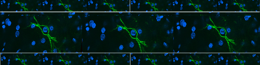
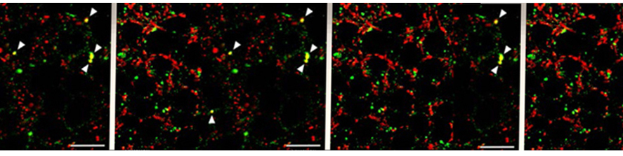
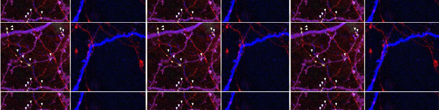
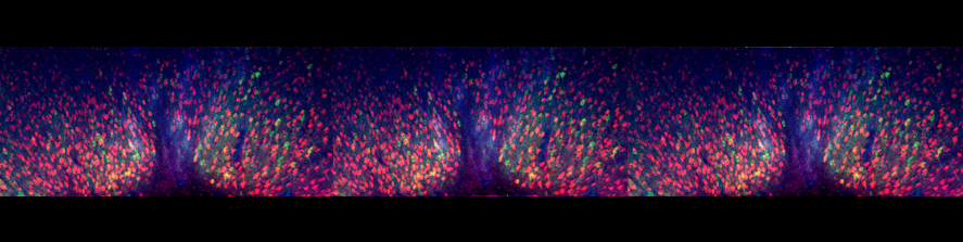
Zeiss LSM900 Confocal Microscope with Airyscan 2. This is a highly integrated, self-contained instrument in which a laser scanning confocal head is interfaced with an inverted Zeiss Axio Observer microscope through a lateral port.
The microscope stand has a table motorized along the x, y and z axes; the objective tower is motorized as well; the microscope is accessorized with DIC optics and a LED camera.
The confocal head uses linear, galvanometric scanners with pinholes that can be modified continuously, PMT photomultipliers, a high sensitivity GaAsP detector and the innovative Airyscan 2 detector.
The new Airyscan technology (which can be exemplified as an array of pinholes) allows a true super resolution, 1.7 times beyond the diffraction limit, as well as a considerable improvement of the signal to noise ratio.
The laser station is composed of 4 solid-state lasers, with emission lines at 405 nm, 488 nm, 561 nm and 640 nm.
The imaging system is controlled by the ZEN 3.0 software, running on a Windows PC with 192 GB RAM. Zen is a highly versatile software including multichannel visualization options, colocalization routines and possibility of quantitative image analysis. Image acquisition menus can be conveniently configured and memorized following users’ requirements.
Noticeably, image acquisition parameters can be reproduced faithfully by simply opening an image file with the Zen3 proprietary extension and using the “apply” routine.
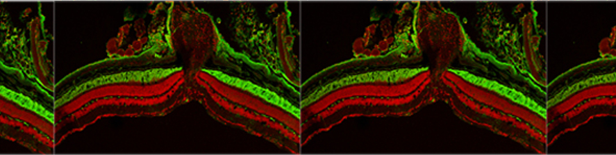
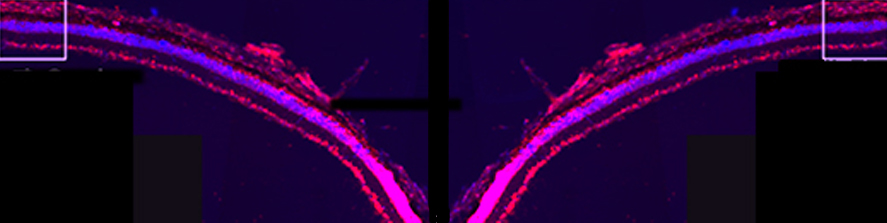
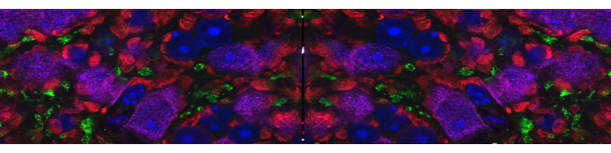
The LSM900 confocal microscope is highly flexible, being adequate for in vitro and ex vivo studies, allowing high definition imaging of specimens containing up to 4 fluorophores, plus a transmitted image with DIC.
ZEN 3.0 software also runs the Zeiss Apotome microscope (part of the BioEnable platform): this allows fast transition between the two instruments as well as high portability of the images obtained by confocal and wide field optical conditions.